






Probe-Style B-Ultrasound: Which Departments It Fits and How to Choose a Wireless Mini Probe
- 2026-11-13
- 29
- Guangzhou Sonostar Technologies Co., Limited
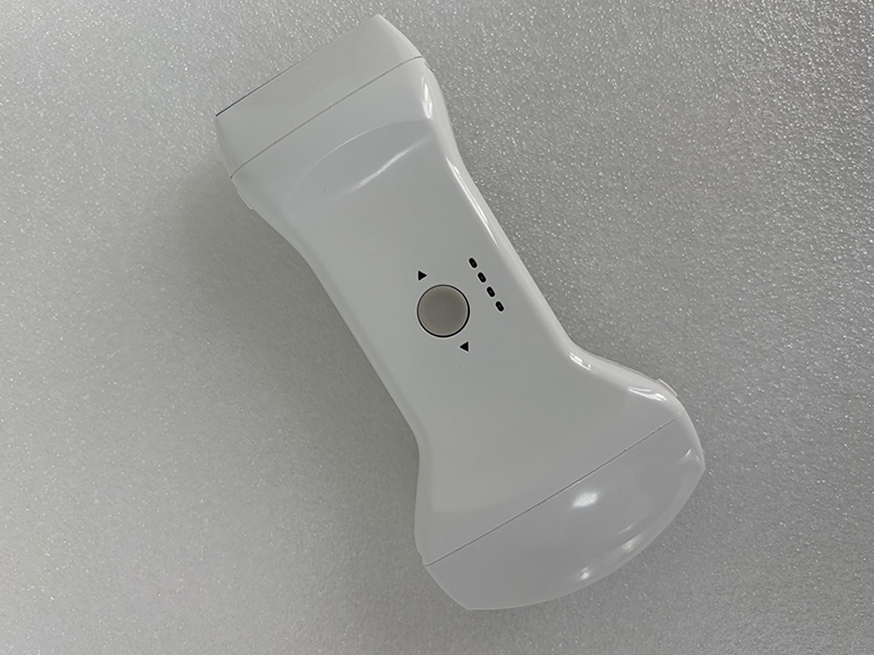
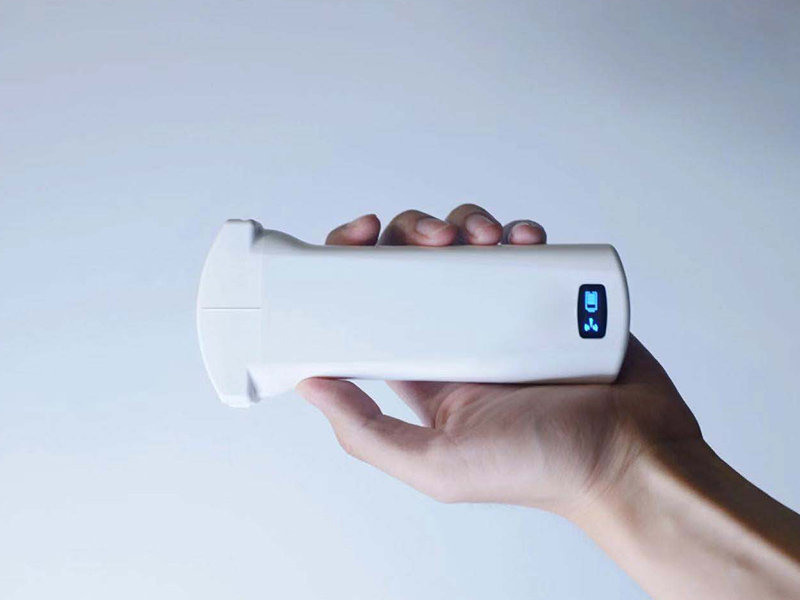
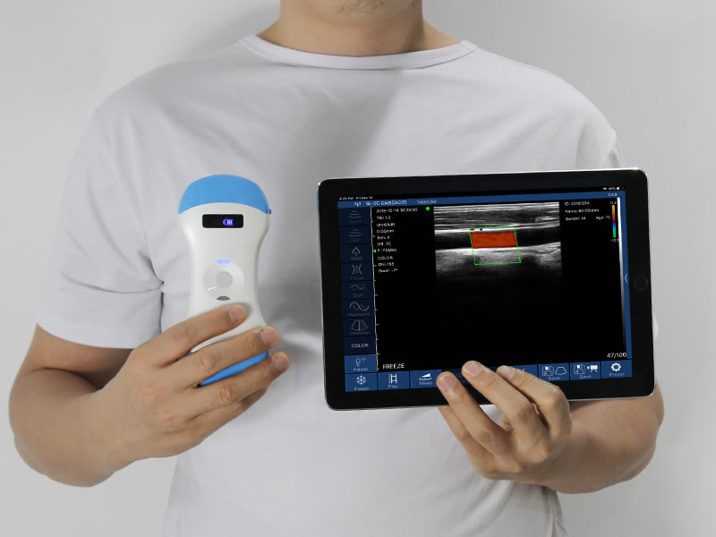
A probe-style B-ultrasound puts the scanner inside the transducer: the beamformer, the battery and the Wi-Fi link sit in the head, and the image appears on a phone or tablet. That changes the adoption question. Nobody asks whether a department can justify a trolley; they ask which single problem the probe solves that day. The answers cluster into four groups, and each group points to a different probe format.
Which departments adopt one, and why
Where the needle goes. Anaesthesia uses it for internal jugular and subclavian lines, cervical and brachial plexus blocks, and spinal access. Pain clinics use it for joint and nerve injections, radiofrequency procedures and soft-tissue release. Nursing uses it for PICC and CVC placement. Nephrology uses it on the fistula: stenosis, thrombosis and calcification before cannulation, and guidance when a new access is created.
Where fluid hides. ICU and emergency look for free fluid, pleural effusion, pneumothorax and pericardial effusion; respiratory medicine assesses effusions and guides drainage; cardiology uses a phased array for cardiac function and volume status. The probe earns its place because the patient does not have to be moved to find out.
Where screening happens. Urology checks residual urine and the prostate; obstetrics and gynaecology cover follicle tracking, endometrial thickness, IUD position and basic fetal assessment; neonatology screens hips and cranial views; health-check centres use it on thyroid, breast, prostate and fatty liver.
Outside the hospital. Village clinics, family doctors on home visits, nursing homes, ambulance crews, sports medical teams and veterinary practices — settings where a trolley is simply not an option.
In every case the probe is a second pair of eyes for the clinician holding it, not a replacement for the ultrasound department. Deep diagnostic work still belongs to the cart and the sonographer, and a clinic that presents the probe as a cart substitute will lose the argument in the opening meeting.
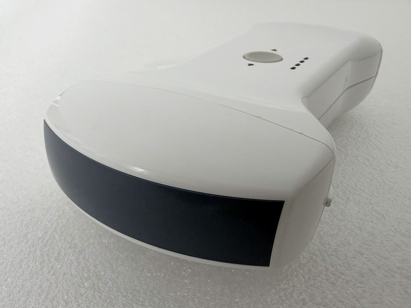
Selecting a probe-style B-ultrasound by format
Start from the view the department takes every week, then pick the head that produces it.
Convex covers abdominal organs, obstetrics, bladder and free fluid — the default for ICU, emergency and village clinics.
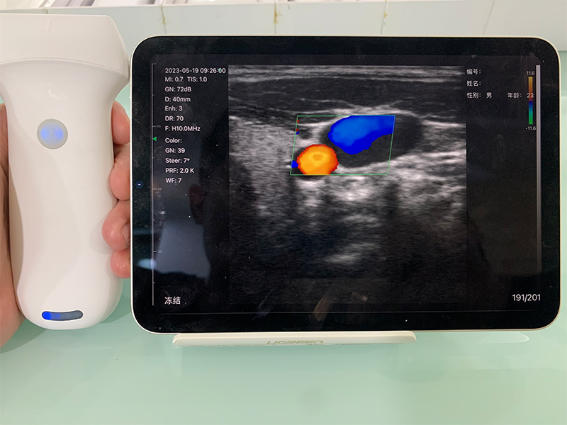
Linear high-frequency covers vessels, thyroid, breast, musculoskeletal work, nerve blocks and PICC.
Ultra-high-frequency goes shallower: skin, tendon, superficial nerve, some veterinary use.
Phased array gives the sector shape that a cardiac window needs through the rib spaces.
Endocavitary and biplane reach transrectal, prostate and gynaecology work; the biplane head is the format for probe-guided puncture because it holds the needle in two planes at once.
Finger-tip heads work in tight spaces, intraoperatively and for veterinary pregnancy checks.
A 3-in-1 head carries two formats in one housing, which is the honest choice for a clinic that cannot buy three probes.
Two specifications matter beyond format. Channel count — 64 on the current models — shows up in image sharpness rather than in the catalogue. And the app platform decides the device budget: the 3D software runs on iOS, so confirm which platforms the 2D models support before standardising on tablets.
FAQ
Can a probe-style B-ultrasound replace the department's ultrasound request?
No. It answers a bedside question in under a minute. Formal studies, measurements and signed reports stay with the ultrasound department, which is also why adoption tends to raise referral quality rather than volume.
Do clinicians need formal ultrasound training?
For screening and needle guidance, a short structured course plus supervised cases is enough to start; certification requirements vary by country and by procedure. Cardiac and obstetric diagnostic work is a different level of training.
Does it work without hospital Wi-Fi?
Yes. The probe broadcasts its own access point, so it runs in an ambulance or a village clinic with no network at all.
How long does a charge last?
Current 2D models run around three hours per charge and recharge in two to three hours over USB; the 3D convex model uses two 2.200 mAh cells at roughly 1.5 hours each, so a spare covers a full day.
Guangzhou SonoStar Information Technology Co., Ltd. is a national high-tech enterprise specializing in miniaturized, intelligent ultrasound. We develop and manufacture handheld probe-style intelligent ultrasound imaging diagnostic devices, wearable B-mode ultrasound and other portable systems, supported by long-term industry–academia–research collaboration. CE- and FDA-registered, our products reach more than 100 countries and regions and serve clinical care, primary and emergency medicine, aesthetic medicine and veterinary practice — advancing accessible ultrasound for everyday care. Contact: Ms. Lin, +86 181 2686 2475.



 Home
 Product
 News
 Contact