How Mini ultrasound is ultilized in the diagnosis of inguinal lymph node?
- 2023-03-03
- 4209
- Guangzhou Sonostar Technologies Co., Limited
- by Naroopa N, Registered Diagnostic Medical Sonographer From USA
Case
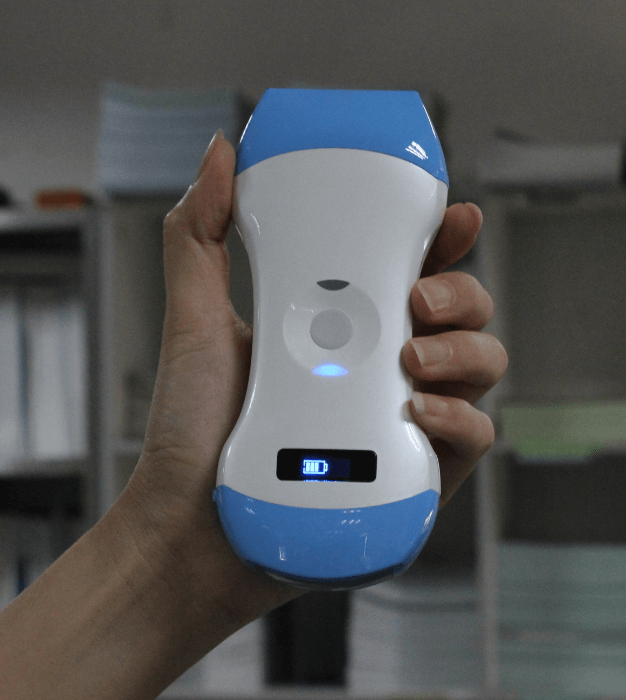
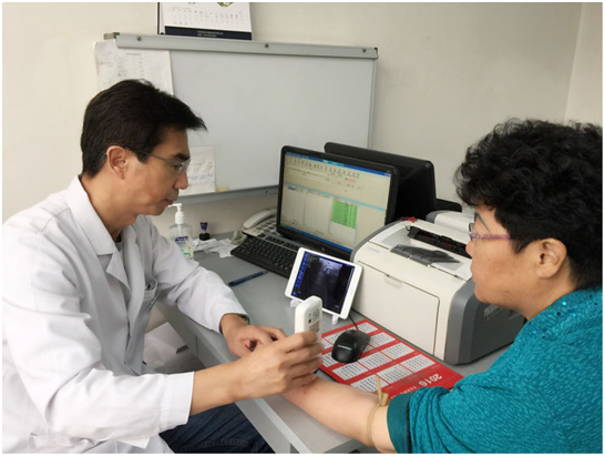
The patient is a 70-year-old woman. The patient is having right leg swelling and pain in the upper leg medial area. Ultrasound scan is performed using the Mini Ultrasound . The deep femoral artery and vein are observed in the sagittal plane clearly. Upon scanning the patient to rule out DVT (deep vein thrombosis) the deep veins are clearly observed and close with compression, as shown below.
Color doppler is applied and demonstrated a beautiful doppler flow in the deep veins and the deep arteries with pulsed wave option. Pulsed wave shows a clear triphasic wave form in the femoral artery.
Upon augmentation, the femoral vein is showing filling in blue doppler color and the difference in the waveform is clearly displayed when augmentation occurs.
Conclusion
Upon scanning the area of pain; two inguinal lymph nodes areas seen on the right side. Impression: There was no evidence of deep vein thrombus or arterial obstruction. The patient has numerous inguinal lymph nodes that were measured and will be monitored.


 Home
Home Product
 News
News Contact